Glycosaminoglycan Array

10609

From: $1,080.00

Glycosaminoglycans (GAGs) are a class of long, linear, and structurally different polysaccharides defined by specific monosaccharides, sulfation modifications, and chemical linkages. The glycosaminoglycan array features 47 distinct GAG structures varying in length, degree of sulfation, and disaccharide sequence. It allows researchers to define GAG-binding specificities for various biological samples, such as proteins, antibodies, cells, cell lysate, serum, vesicles, bacteria, or viral particles. Each array contains 8 or 16 identical subarrays, enabling the simultaneous analysis of multiple samples. The glycosaminoglycan array provides high-throughput and reliable glycan-binding information with a simple assay format that only requires a small sample volume. It can be customized to meet individual client needs. Assay services are available upon request.

SKU: 10609 Category:
Description
Structures
Examples
Citations
Document

Description

Glycosaminoglycans (GAGs), also known as mucopolysaccharides, are a class of long and linear polysaccharides. There are four major classes of GAGs: hyaluronic acid (HA), heparan sulfate/heparin (HS/Hep), chondroitin sulfate/dermatan sulfate (CS/DS), and keratan sulfate (KS). They are structurally different polysaccharides defined by specific monosaccharides, sulfation modifications, and chemical linkages.

GAGs are ubiquitously found on the mammalian cell surface and in the extracellular matrix. They maintain cell hydration and provide structural support for connective tissues. They are also critical regulators in cell signaling pathways. However, abnormally expressed GAGs have been linked to various diseases. For example, alterations in GAG sulfation patterns have been associated with poor prognosis of breast, ovarian, colorectal, prostate, and gastric cancers. However, it is not fully understood to what extent the degree of GAG sulfation can promote or inhibit cancer progression. One of the hallmarks of Alzheimer’s disease is the presence of extracellular plaques composed of fibrillated amyloid-beta (Aβ) protein. Sulfated GAGs act as pathological chaperones to assist Ab aggregation. However, GAGs of a unique N-sulfation pattern can reduce Aβ protein aggregation. GAGs are often targeted by microbial pathogens to enter the cell and establish infections. Many microbial pathogens hijack the host biological process of GAG synthesis to subvert immunity and promote disease. Therefore, identifying and understanding the differences between GAGs expressed in normal and pathological conditions is vital for understanding the pathogenesis of these diseases.

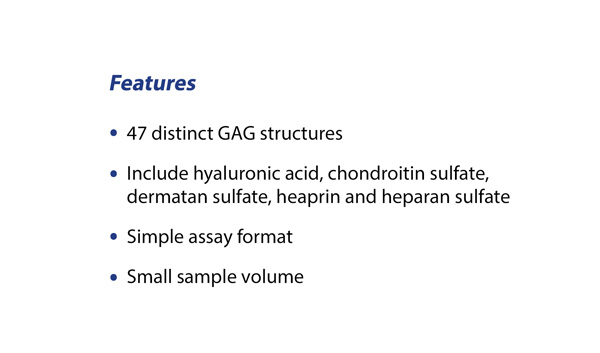
ZBiotech has developed a robust microarray platform that allows researchers to define GAG-binding specificities for various biological samples, such as proteins, antibodies, cells, cell lysate, serum, vesicles, bacteria, or viral particles. The glycosaminoglycan array features 47 distinct GAG structures varying in length, degree of sulfation, and disaccharide sequence. Each array contains 8 or 16 identical subarrays, enabling the simultaneous analysis of multiple samples. The glycosaminoglycan array provides high-throughput and reliable glycan-binding information with a simple assay format that only requires a small sample volume. It can be customized to meet individual client needs. Assay services are available upon request.

Features

  • Unrivaled sensitivity and specificity;
  • Simple assay format;
  • Small sample volume;
  • Customizable (select GAGs for a specific microarray format)
  • Assay service available;

Applications

  • Evaluate binding specificities of GAG-interacting proteins;
  • Evaluate binding specificities of GAG-interacting antibodies;
  • Study virus – GAG interactions;
  • Study bacteria – GAG glycan interactions;
  • Study vesicle – GAG glycan interactions;
  • Study cell – GAG glycan interactions;

Structures

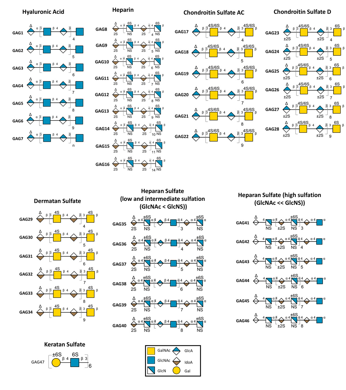
List of glycosaminoglycan structures on the array (download the PDF)

ID Name Structure and Molecular Weight
Hyaluronic Acid (HA) GAG1 Hyaluronic Acid dp10 (HA10) ∆GlcAβ1,3 [GlcNAcβ1,4 GlcAβ1,3]4 GlcNAc, Mw 1,950 Da
GAG2 Hyaluronic Acid dp12 (HA12) ∆GlcAβ1,3 [GlcNAcβ1,4 GlcAβ1,3]5 GlcNAc, Mw 2,350 Da
GAG3 Hyaluronic Acid dp14 (HA14) ∆GlcAβ1,3 [GlcNAcβ1,4 GlcAβ1,3]6 GlcNAc, Mw 2,700 Da
GAG4 Hyaluronic Acid dp16 (HA16) ∆GlcAβ1,3 [GlcNAcβ1,4 GlcAβ1,3]7 GlcNAc, Mw 3,150 Da
GAG5 Hyaluronic Acid dp18 (HA18) ∆GlcAβ1,3 [GlcNAcβ1,4 GlcAβ1,3]8 GlcNAc, Mw 3,650 Da
GAG6 Hyaluronic Acid dp20 (HA20) ∆GlcAβ1,3 [GlcNAcβ1,4 GlcAβ1,3]9 GlcNAc, Mw 3,900 Da
GAG7 Hyaluronic Acid Polymer (HA93) ∆GlcAβ1,3 [GlcNAcβ1,4 GlcAβ1,3]n GlcNAc, Mw 93 kDa
Heparin GAG8 Heparin dp10 (H10) ∆UA,2S – GlcNS,6S– [IdoUA,2S – GlcNS,6S]4, Mw 3,000
GAG9 Heparin dp12 (H12) ∆UA,2S – GlcNS,6S– [IdoUA,2S – GlcNS,6S]5, Mw 3,550
GAG10 Heparin dp14 (H14) ∆UA,2S – GlcNS,6S– [IdoUA,2S – GlcNS,6S]6, Mw 4,100
GAG11 Heparin dp16 (H16) ∆UA,2S – GlcNS,6S– [IdoUA,2S – GlcNS,6S]7, Mw 4,650
GAG12 Heparin dp18 (H18) ∆UA,2S – GlcNS,6S– [IdoUA,2S – GlcNS,6S]8, Mw 5,200
GAG13 Heparin dp20 (H20) ∆UA,2S – GlcNS,6S– [IdoUA,2S – GlcNS,6S]9, Mw 5,750
GAG14 Heparin dp22 (H22) ∆UA,2S – GlcNS,6S– [IdoUA,2S – GlcNS,6S]10, Mw 6,300
GAG15 Heparin dp24 (H24) ∆UA,2S – GlcNS,6S– [IdoUA,2S – GlcNS,6S]11, Mw 6,850
GAG16 Heparin dp30 (H30) ∆UA,2S – GlcNS,6S– [IdoUA,2S – GlcNS,6S]14, Mw 9,000
Chondroitin Sulfate (CS) GAG17 Chondroitin Sulphate Oligosaccharide dp10 (CSO10) ∆UA – [GalNAc,6S or 4S – GlcA]4 – GalNAc,6S or 4S, Mw 2,480
GAG18 Chondroitin Sulphate Oligosaccharide dp12 (CSO12) ∆UA – [GalNAc,6S or 4S – GlcA]5 – GalNAc,6S or 4S, Mw 2,976
GAG19 Chondroitin Sulphate Oligosaccharide dp14 (CSO14) ∆UA – [GalNAc,6S or 4S – GlcA]6 – GalNAc,6S or 4S, Mw 3,472
GAG20 Chondroitin Sulphate Oligosaccharide dp16 (CSO16) ∆UA – [GalNAc,6S or 4S – GlcA]7 – GalNAc,6S or 4S, Mw 3,968
GAG21 Chondroitin Sulphate Oligosaccharide dp18 (CSO18) ∆UA – [GalNAc,6S or 4S – GlcA]8 – GalNAc,6S or 4S, Mw 4,464
GAG22 Chondroitin Sulphate Oligosaccharide dp20 (CSO20) ∆UA – [GalNAc,6S or 4S – GlcA]9 – GalNAc,6S or 4S, Mw 4,960
GAG23 Chondroitin Sulphate D Oligosaccharide dp10 (CSDO10) ∆UA – [GalNAc,6S or 4S – GlcA +/- 2S]4 – GalNAc,6S, Mw 2,480
GAG24 Chondroitin Sulphate D Oligosaccharide dp12 (CSDO12) ∆UA – [GalNAc,6S or 4S – GlcA +/- 2S]5 – GalNAc,6S, Mw 2,976
GAG25 Chondroitin Sulphate D Oligosaccharide dp14 (CSDO14) ∆UA – [GalNAc,6S or 4S – GlcA +/- 2S]6 – GalNAc,6S, Mw 3,472
GAG26 Chondroitin Sulphate D Oligosaccharide dp16 (CSDO16) ∆UA – [GalNAc,6S or 4S – GlcA +/- 2S]7 – GalNAc,6S, Mw 3,968
GAG27 Chondroitin Sulphate D Oligosaccharide dp18 (CSDO18) ∆UA – [GalNAc,6S or 4S – GlcA +/- 2S]8 – GalNAc,6S, Mw 4,464
GAG28 Chondroitin Sulphate D Oligosaccharide dp20 (CSDO20) ∆UA – [GalNAc,6S or 4S – GlcA +/- 2S]9 – GalNAc,6S, Mw 4,960
Dermatan Sulfate (DS) GAG29 Dermatan Sulphate dp10 (DS10) ∆UAβ1,3 – GalNAc,4S – [IdoA – GalNAc,4S]4, Mw 2,480
GAG30 Dermatan Sulphate dp12 (DS12) ∆UAβ1,3 – GalNAc,4S – [IdoA – GalNAc,4S]5, Mw 2,976
GAG31 Dermatan Sulphate dp14 (DS14) ∆UAβ1,3 – GalNAc,4S – [IdoA – GalNAc,4S]6, Mw 3,472
GAG32 Dermatan Sulphate dp16 (DS16) ∆UAβ1,3 – GalNAc,4S – [IdoA – GalNAc,4S]7, Mw 3,968
GAG33 Dermatan Sulphate dp18 (DS18) ∆UAβ1,3 – GalNAc,4S – [IdoA – GalNAc,4S]8, Mw 4,464
GAG34 Dermatan Sulphate dp20 (DS20) ∆UAβ1,3 – GalNAc,4S – [IdoA – GalNAc,4S]9, Mw 4,960
Heparan Sulfate (HS) GAG35 Heparan Sulphate Oligosaccharide dp10 (Hep I, low and intermediate sulphation) ∆UA2S – GlcNS – [GlcA – GlcNAc]3 – IdoA – GlcNS, Mw 2,800
GAG36 Heparan Sulphate Oligosaccharide dp12 (Hep I, low and intermediate sulphation) ∆UA2S – GlcNS – [GlcA – GlcNAc]4 – IdoA – GlcNS, Mw 3,500
GAG37 Heparan Sulphate Oligosaccharide dp14 (Hep I, low and intermediate sulphation) ∆UA2S – GlcNS – [GlcA – GlcNAc]5 – IdoA – GlcNS, Mw 4,000
GAG38 Heparan Sulphate Oligosaccharide dp16 (Hep I, low and intermediate sulphation) ∆UA2S – GlcNS – [GlcA – GlcNAc]6 – IdoA – GlcNS, Mw 4,400
GAG39 Heparan Sulphate Oligosaccharide dp18 (Hep I, low and intermediate sulphation) ∆UA2S – GlcNS – [GlcA – GlcNAc]7 – IdoA – GlcNS, Mw 5,000
GAG40 Heparan Sulphate Oligosaccharide dp20 (Hep I, low and intermediate sulphation) ∆UA2S – GlcNS – [GlcA – GlcNAc]8 – IdoA – GlcNS, Mw 5,400
GAG41 Heparan Sulphate Oligosaccharide dp10 (Hep III, high sulphation) ∆UA – GlcNS – [IdoA +/- 2S – GlcNS]3 – IdoA – GlcNAc, Mw 2,800
GAG42 Heparan Sulphate Oligosaccharide dp12 (Hep III, high sulphation) ∆UA – GlcNS – [IdoA +/- 2S – GlcNS]4 – IdoA – GlcNAc, Mw 3,500
GAG43 Heparan Sulphate Oligosaccharide dp14 (Hep III, high sulphation) ∆UA – GlcNS – [IdoA +/- 2S – GlcNS]5 – IdoA – GlcNAc, Mw 4,200
GAG44 Heparan Sulphate Oligosaccharide dp16 (Hep III, high sulphation) ∆UA – GlcNS – [IdoA +/- 2S – GlcNS]6 – IdoA – GlcNAc, Mw 4,800
GAG45 Heparan Sulphate Oligosaccharide dp18 (Hep III, high sulphation) ∆UA – GlcNS – [IdoA +/- 2S – GlcNS]7 – IdoA – GlcNAc, Mw 5,500
GAG46 Heparan Sulphate Oligosaccharide dp20 (Hep III, high sulphation) ∆UA – GlcNS – [IdoA +/- 2S – GlcNS]8 – IdoA – GlcNAc, Mw 6,200
Keratan Sulfate (KS) GAG47 Keratan Sulfate Oligosaccharide [Gal +/- 6S – GlcNAc, 6S]6, Mw 3,000

Hep I: Heparinase I; Hep III: Heparinase III

Examples

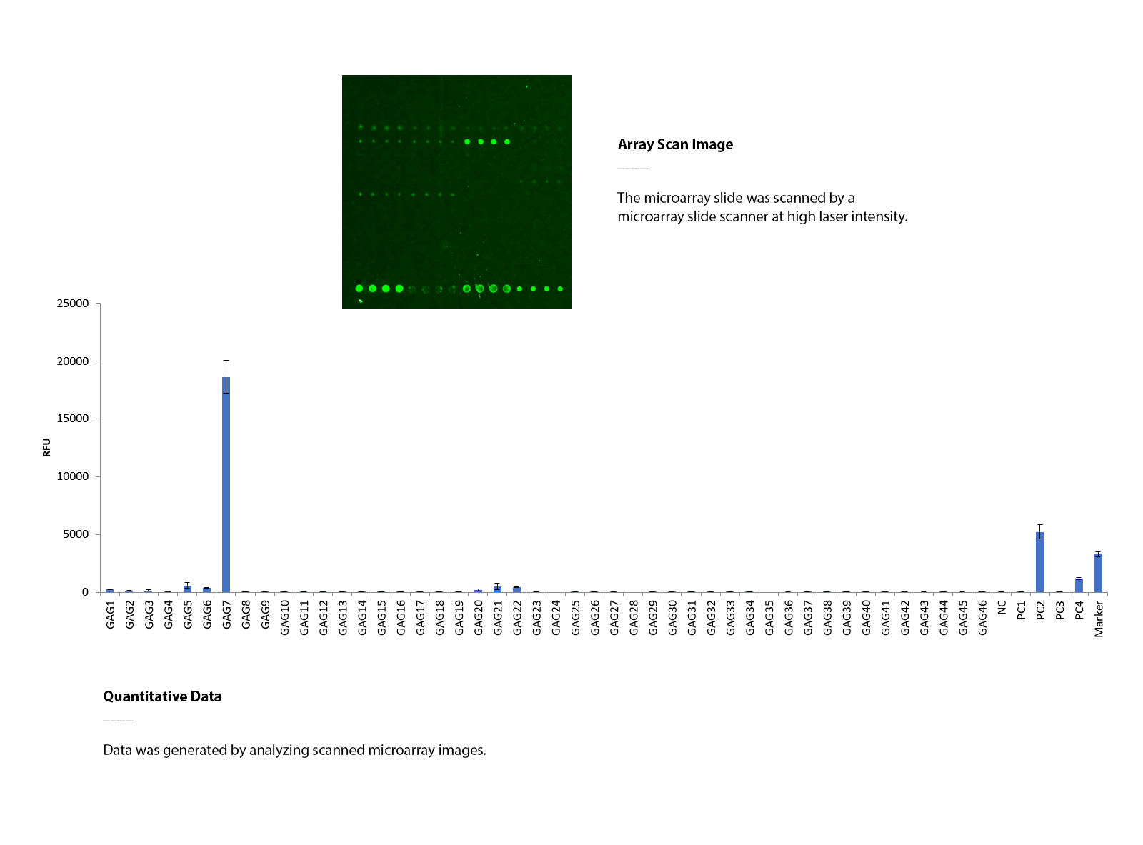
Using glycosaminoglycan array to determine the binding specificity of mouse CD44

The glycosaminoglycan array was assayed with mouse CD44 hFc (5 μg/mL), followed by an anti-human IgG antibody (Cy3). The array was scanned with a microarray scanner at 532nm wavelength. Positive control showed binding signals as expected. CD44 interacts with hyaluronic acid (GAG7).

Document

List of glycosaminoglycan structures on the array (download the PDF)

Protocol & User Manual (download the manual)