ZBiotech’s Neu5Ac and Neu5Gc N-glycan microarray helps researchers identify a carbohydrate-based biomarker for cancer detection.

A protective surface that bacteria can exploit
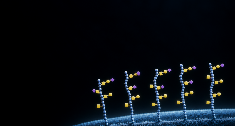
The intestinal lining is covered with mucins: large proteins densely decorated with sugar chains called glycans. These molecules help protect epithelial cells and shape their interactions with microbes. Some pathogens, however, can use this protective surface as a foothold. One example is Salmonella enterica. Its giant adhesion protein, SiiE, can engage MUC1, a mucin anchored to the surface of intestinal cells, and support invasion from the side facing the intestinal lumen.
Earlier research had established that this interaction depends on sialic acids, sugars that often cap mucin glycans. But that left an important question unanswered: Which glycan structures does SiiE recognize, and how does their arrangement on MUC1 affect binding? Researchers at Utrecht University, Leiden University Medical Center, NOVA University Lisbon and TNO addressed this question.
Why the position of a glycan matters
MUC1 contains a variable number of repeating amino acid sequences, known as tandem repeats. Each repeat provides several serine and threonine residues where O-glycans can be attached. Changing the glycan structure, its attachment site or the number of occupied sites changes the surface presented to a binding protein. This arrangement is sometimes described as a “glycan barcode.” The information lies in both the sugars themselves and the pattern in which they are displayed.
For SiiE, that pattern could be particularly important. At approximately 600 kDa, SiiE is an unusually large protein with 53 lectin-like repeat domains. Understanding how it interacts with densely glycosylated MUC1 therefore requires more than a list of individual sugars it can bind.
How the Z Biotech MUC1 Glycopeptide Array 2 helped
The researchers used the Z Biotech MUC1 Glycopeptide Array to compare defined glycan presentations on a common peptide backbone. The array contained 124 synthetic glycopeptides based on the same 23-amino-acid MUC1 sequence, together with a nonglycosylated peptide control. The library covered four glycan classes: core 2, α2,3-sialylT, α2,6-sialyl-T and disialyl-T. Within each class, glycans were attached at defined positions, with different numbers and combinations of occupied sites.
This design allowed the researchers to examine three connected questions: which glycan structures supported binding, which attachment sites were preferred, and how adding more glycans affected the response. Because the peptide sequence remained constant, differences could be interpreted against a shared MUC1 background.
The team tested an enriched fraction of secreted SiiE from bacterial culture supernatant and detected bound SiiE through antibody-based fluorescence. A preparation from bacteria lacking SiiE served as a negative control, while the lectin PNA provided a reference for recognition of exposed galactose-containing motifs.
A binding profile shaped by structure and presentation
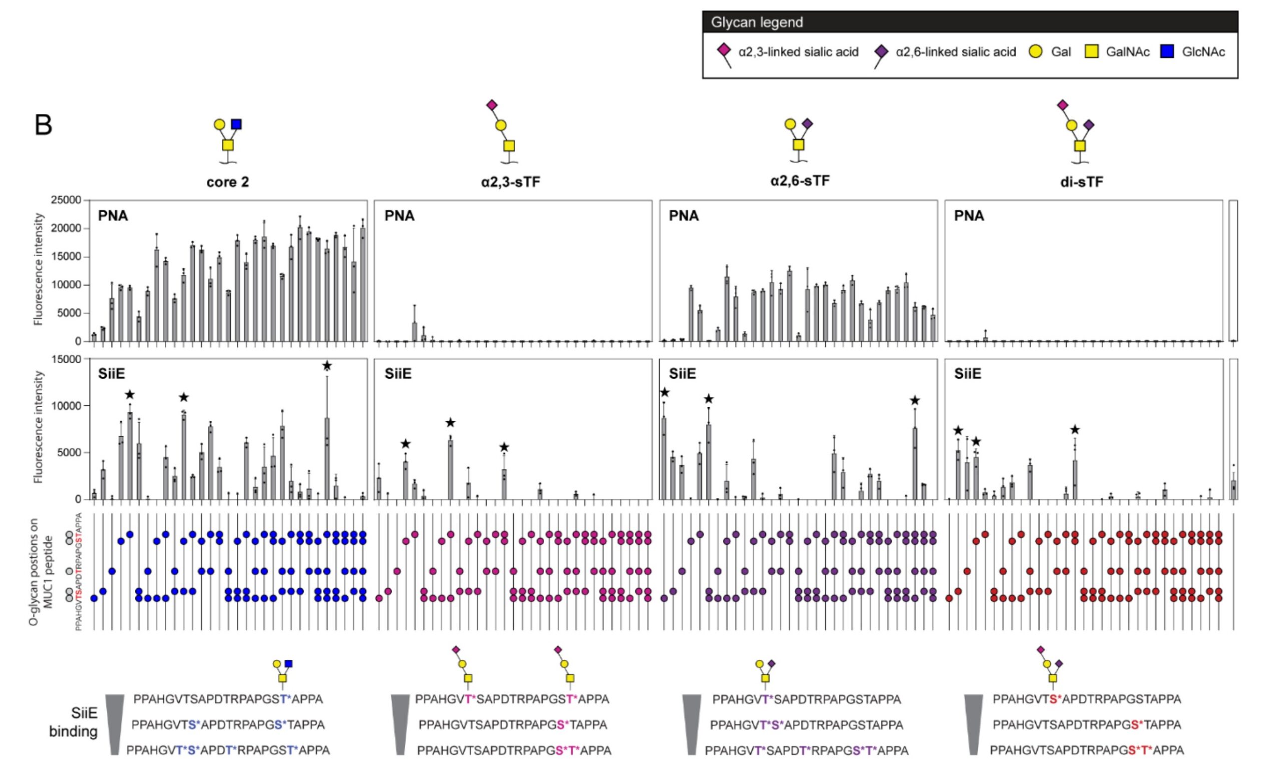
SiiE recognized glycopeptides from all four glycan classes, but its responses varied with glycan structure and arrangement.
| Glycan class What the researchers observed | Glycan class What the researchers observed |
| Core 2 | The strongest SiiE binding signals, consistent with earlier evidence of GlcNAc recognition. |
| α2,3-sialyl-T | Moderate binding that depended strongly on glycan position and tended to decrease as more sites were glycosylated. |
| α2,6-sialyl-T | Position-dependent binding, including recognition of some peptides with higher glycan density. |
| Disialyl-T | Moderate binding, with a preference for peptides carrying fewer glycans. |
The preferred glycan positions also differed among the sialylated classes. Knowing which glycan was present was therefore not enough to predict the binding response; knowing where it was attached added essential information. That is the value of a defined glycopeptide panel. Native mucin preparations contain mixtures of glycoforms that can obscure these comparisons. The array made it possible to examine selected changes in glycan presentation across a consistent peptide sequence

Figure 1. SiiE binding to defined MUC1 glycopeptides on the Z Biotech MUC1 Glycopeptide Array 2. Glycopeptides are grouped by core 2, α2,3-sialyl-T, α2,6-sialyl-T and disialyl-T structures. PNA serves as a control for exposed galactose-containing motifs; SiiE signals show how binding varies with glycan class and presentation. Colored circles indicate glycosylation sites, and stars identify the three highest-binding glycopeptides in each class. Bars represent mean ± SD from three independent biological replicates. Reproduced from Figure 3B of Giesbers et al., bioRxiv (2026), under CC BY 4.0.
Connecting array binding to intestinal invasion
The array findings became especially informative when considered alongside experiments in intestinal cells. Although secreted SiiE recognized several glycan classes on the array, the cell-based experiments identified a more specific requirement for invasion. Removing α2,3-linked sialic acids, or blocking them with MAL II, reduced bacterial invasion to levels comparable to those seen in cells lacking MUC1. Blocking with the α2,6-directed lectin SNA did not produce the same reduction. Glycomics analysis also showed that MUC1 from the intestinal cell model carried prominent α2,3- sialylated Oglycans. Together, these experiments supported a requirement for α2,3-sialylated MUC1 in the invasion pathway studied.
The difference between array binding and cell invasion led to a useful hypothesis. On native MUC1, repeated glycans may allow several SiiE domains to engage together, strengthening attachment through multiple contacts. The authors propose that the arrangement of α2,3-sialylated glycans along MUC1 helps create a favorable surface for this interaction. The precise multivalent arrangement remains to be established.
Experiments with human intestinal tissue added biological context: in the colonic tissue examined, Salmonella was observed close to MUC1 associated with α2,3-sialic acid staining. This supported further investigation of the pathway in the human colon.
What this approach can bring to your research
For researchers studying MUC1 recognition, a binding result often raises further questions. Does a glycan promote recognition or mask a peptide epitope? Does moving the same glycan to another site alter binding? Does increasing glycosylation strengthen or reduce the response?
The MUC1 Glycopeptide Array provides defined comparisons that can help address these questions. Potential applications include:
In the SiiE study, the array supplied a detailed view of glycan recognition that complemented the functional experiments. It helped researchers connect a specific binding pattern to a broader biological question: how a pathogen recognizes the complex, glycosylated surface of its host.
Giesbers, K. C. A. P., et al. (2026). Salmonella SiiE-mediated apical invasion into colonocytes
depends on MUC1 α2,3-linked sialic acids. bioRxiv. https://doi.org/10.64898/2026.07.21.739827
This feature discusses the July 21, 2026 preprint, which had not undergone peer review in the version reviewed here.