A New Clue to How H3N2 Influenza Attaches to Cells

ZBiotechMicrobiologyA New Clue to How H3N2 Influenza Attaches to Cells
Microbiology

A New Clue to How H3N2 Influenza Attaches to Cells

Z Biotech’s Alpha 2,3/2,6 Sialylation Profiling Kit helped researchers show that reduced H3N2 attachment was not accompanied by a detectable change in overall cell-surface sialylation.

Highlights

Product: Alpha 2,3/2,6 Sialylation Profiling Kit
Field:Microbiology / Virology
Study:Cell-surface sialylation analysis

An unexpected role in viral attachment

Before influenza can infect a cell, it must attach to the cell’s surface. Researchers at the University of Vermont discovered that a cellular transport protein, Rab11B, plays an unexpected role in this first step.

Rab11B is closely related to Rab11A, a protein known for helping transport influenza’s genetic material during the later stages of infection. But experiments with clinical isolates collected in 2022 revealed an earlier role for Rab11B: helping H3N2 influenza attach to cells.

When the researchers reduced Rab11B expression, H3N2 attachment to A549 lung cells fell by approximately 50%. The H1N1 isolate tested did not show the same attachment defect. Further experiments linked the H3N2 effect to hemagglutinin (HA), the viral protein responsible for recognizing host cells.


Could a change in surface sugars explain the result?

Influenza HA recognizes sialic acids on the cell surface. This raised a straightforward question: Did reducing Rab11B simply leave fewer sialic acids available for the virus to bind?

Answering that question was essential. A broad reduction in surface sialylation could explain the attachment defect without requiring a more specific role for Rab11B.

The researchers therefore needed to compare the two major sialic acid linkages, α2,3 and α2,6, across control cells and cells depleted of Rab11A or Rab11B.


How the Z Biotech kit helped

The team used a flow cytometry workflow adapted from the Z Biotech Alpha 2,3/2,6 Sialylation Profiling Kit protocol. They stained cells with three lectins:

  • SNA, which recognizes α2,6-linked sialic acids.
  • MAL I and MAL II, which recognize different glycan contexts containing α2,3-linked
    sialic acids.

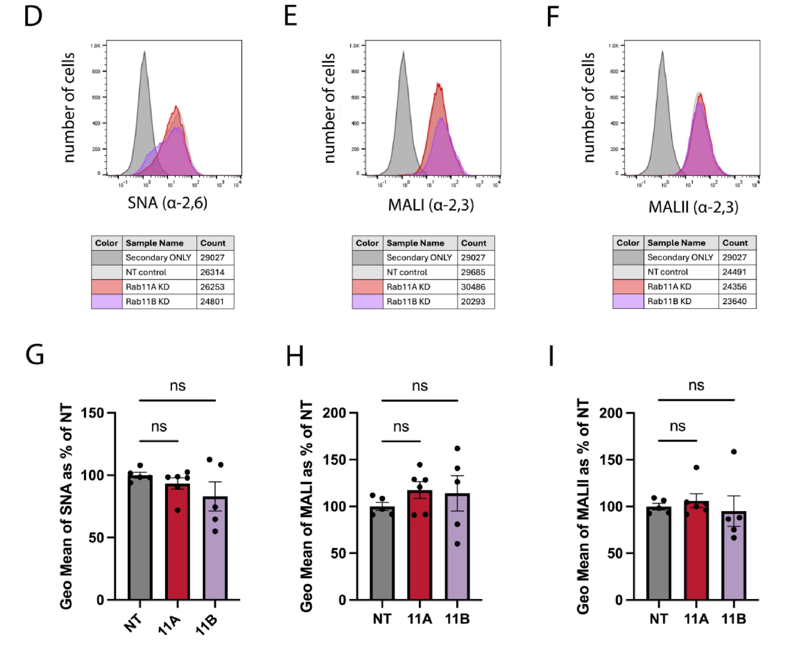
Fluorescent detection allowed the team to compare lectin binding between cell populations. A viability stain identified live cells, while a secondary-only control established background fluorescence. None of the three lectins showed a statistically significant change after Rab11B depletion. The assay therefore provided no evidence of a broad reduction in the surface sialylation detected by these probes.

The profiling results helped the researchers narrow their interpretation. H3N2 attachment had decreased, but the overall sialylation profile measured by the lectins had not. Combined with the other experiments, this supported a more specific hypothesis: Rab11B may help deliver particular sialylated proteins to the cell surface that recent H3N2 viruses use for attachment. The study did not identify those proteins. Instead, the kit supplied an important piece of evidence that helped guide the next stage of receptor research.


Figure 3. Cell-surface sialylation analysis of A549 cells following Rab11A or Rab11B depletion. Panels DF show representative flow cytometry profiles for SNA, MAL I and MAL II; panels G–I summarize normalized fluorescence. No significant change was detected after Rab11B depletion with any of the three lectins. Reproduced from Figure 8D-I of Turner et al., Journal of Virology 100(6), e02111-25 (2026), under CC BY 4.0.


What the kit can do for your research

Changes in cell behavior can raise an important question: has surface sialylation changed too? The Z Biotech Alpha 2,3/2,6 Sialylation Profiling Kit helps researchers compare linkage-associated lectin staining through a flow cytometry workflow. Applications include:

  • Investigating viral attachment and the contribution of cell-surface sialylation.
  • Evaluating glycoengineered or gene-edited cells before downstream
    experiments.
  • Monitoring treatment effects, including changes following sialyltransferase or
    neuraminidase inhibition.
  • Tracking sialylation changes during differentiation, immune activation or cancer research.

In this study, the kit helped researchers evaluate an alternative explanation and build a stronger case for investigating specific H3N2 attachment factors.


Reference

Turner, A. H., et al. (2026). Rab11B is required for binding and entry of recent H3N2, but not H1N1,influenza A isolates. Journal of Virology, 100(6), e02111-25. https://doi.org/10.1128/jvi.02111-25