Z Biotech’s N-Glycan and O-Glycan Arrays helped researchers uncover striking differences in receptor recognition among H5N1 influenza variants, including severely diminished glycan binding in variants identified during a human infection.

Understanding how H5N1 recognizes host cells
Influenza viruses initiate infection through hemagglutinin (HA), a surface protein that recognizes sialic acid-containing glycans on host cells. Avian influenza viruses generally prefer α2,3-linked sialic acids, while human-adapted influenza viruses often recognize α2,6-linked sialic acids. Understanding these binding preferences is important for investigating viral host adaptation and potential transmission between species.
In 2024, a severe human H5N1 infection in British Columbia, Canada, raised questions about two HA mutations, E190D and Q226H, detected in the patient’s viral samples. Could these mutations shift the virus toward recognizing human-type receptors?
Researchers at the University of British Columbia investigated this question by comparing HA proteins from three H5N1 strains: A/Vietnam/1203/2004 (VN04), A/Michigan/90/2024 (MI24), and A/British Columbia/PHL-2032/2024 (BC24). Their findings revealed an unexpected result.
How Z Biotech’s Glycan Arrays helped
To characterize receptor recognition, the researchers used Z Biotech’s N-Glycan and O-Glycan Arrays to examine HA binding across structurally diverse glycans, including α2,3-linked and α2,6- linked sialosides. The two complementary arrays allowed the researchers to compare binding preferences across different glycan structures and identify how individual HA variants responded to specific glycan features.

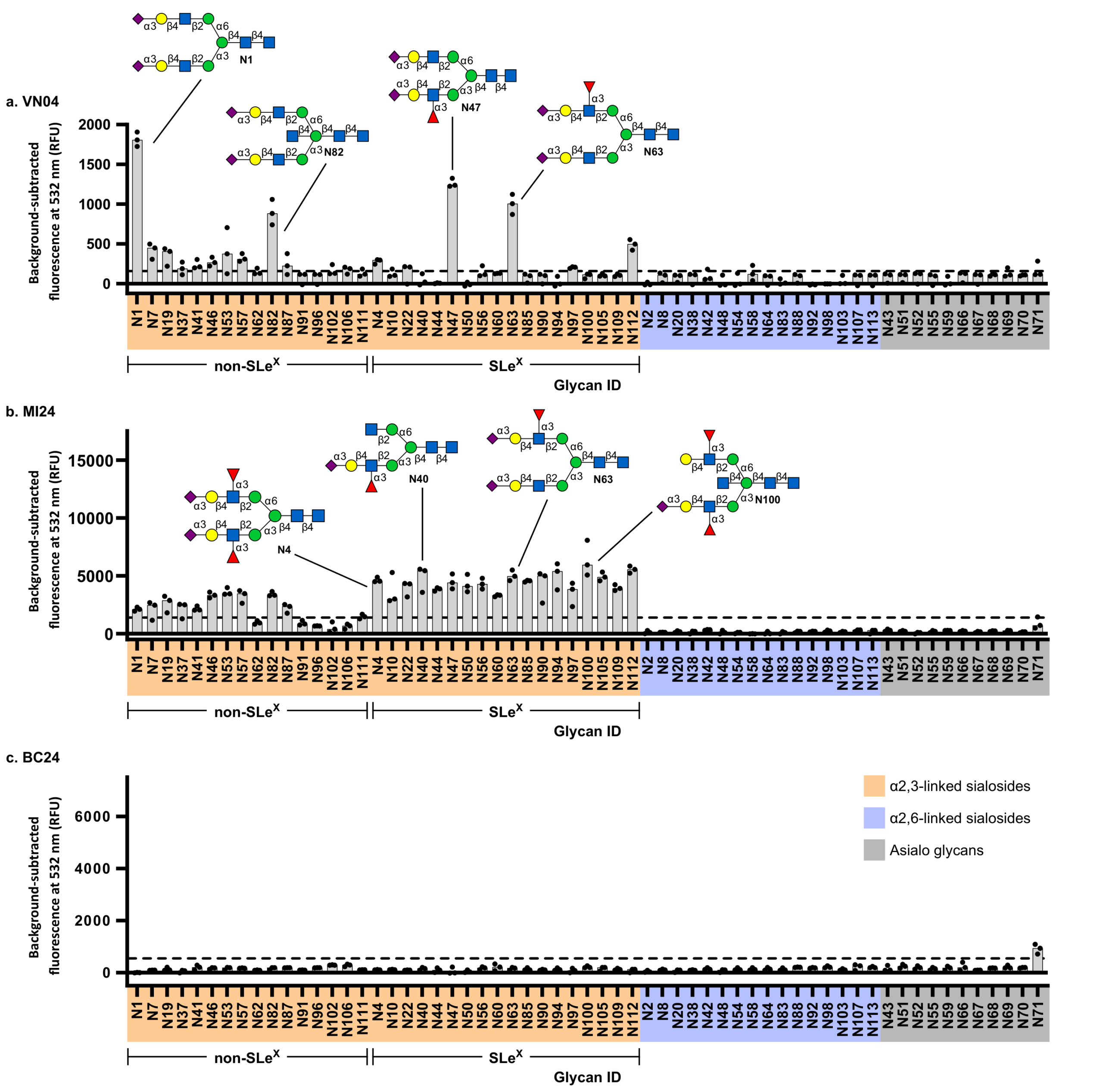
Figure 1. H5N1 hemagglutinin binding profiles revealed by the Z Biotech N-Glycan Array. The NGlycan Array was used to characterize the glycan-binding preferences of three H5N1 hemagglutinin (HA) variants: VN04, MI24, and BC24. VN04 primarily recognized α2,3-linked sialosides, while MI24 exhibited stronger and broader binding, particularly to fucosylated glycans containing sialyl Lewis X (sLeX). In contrast, BC24 showed no significant binding to the tested sialosides above the study’s detection threshold. Orange, α2,3-linked sialosides; blue, α2,6-linked sialosides; gray, asialo glycans. Fluorescence signals were background-subtracted, with three replicate spots per glycan. The dashed line indicates a threshold of 50- fold background signal. Source: Ni et al., Nature Communications (2026), Figure 3.

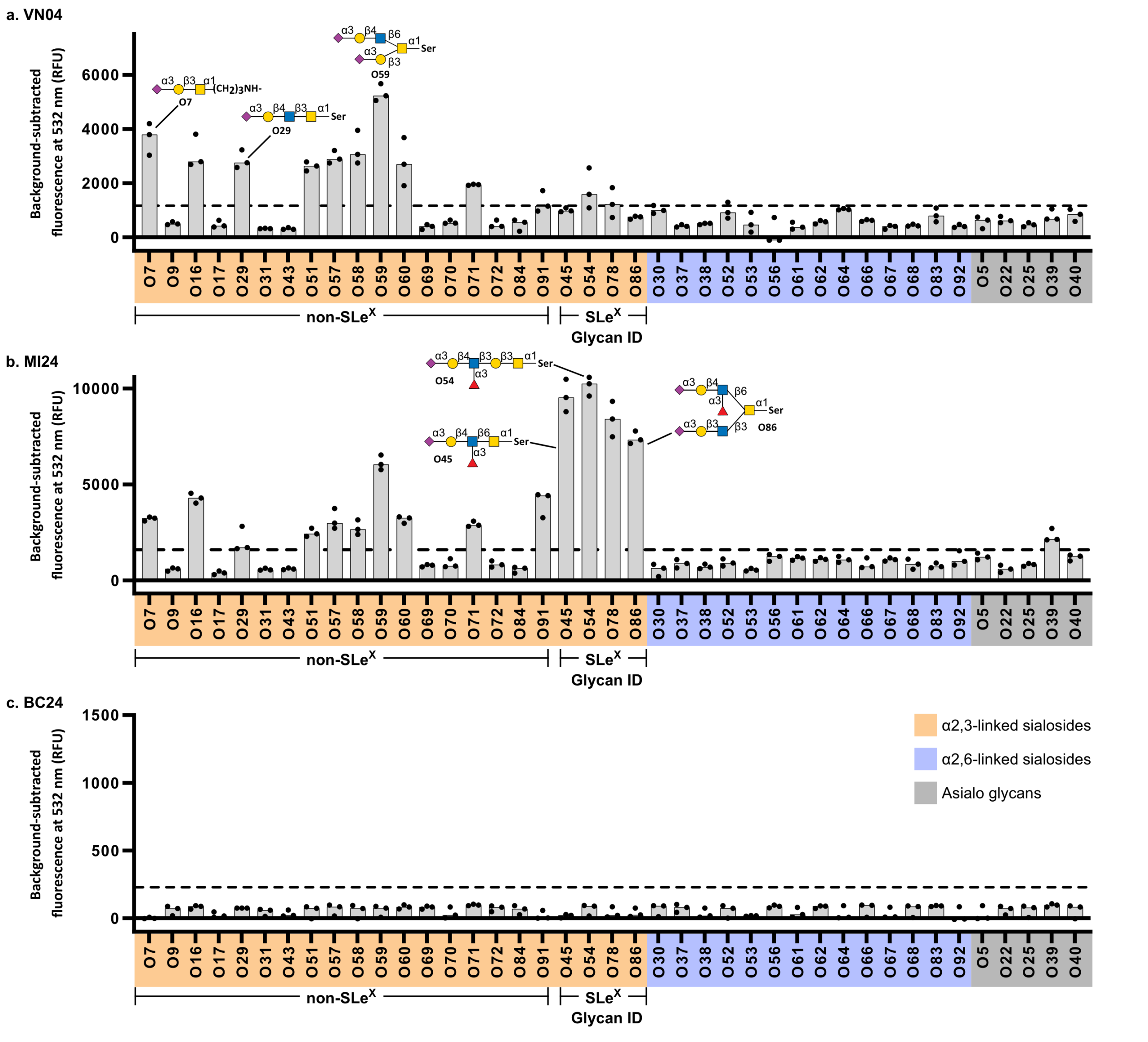
Figure 2. Complementary characterization of H5N1 hemagglutinin receptor specificity using the Z Biotech O-Glycan Array.The O-Glycan Array revealed distinct recognition patterns among VN04, MI24, and BC24 HA variants. VN04 and MI24 preferentially recognized α2,3-linked sialylated O-glycans, with MI24 displaying broader recognition of sialyl Lewis X (sLeX)-containing structures. BC24 showed no detectable binding to the tested sialosides above the study’s threshold, consistent with the N-Glycan Array findings. Orange, α2,3-linked sialosides; blue, α2,6-linked sialosides; gray, asialo glycans. Fluorescence signals were background-subtracted, with three replicate spots per glycan. The dashed line indicates a threshold of 50-fold background signal. Source: Ni et al., Nature Communications (2026), Figure 4
The results revealed three distinct binding profiles:
Importantly, the BC24 variant did not demonstrate the anticipated shift toward human-type α2,6- linked receptors. Instead, it exhibited a striking reduction in detectable glycan binding. By profiling many glycan structures in parallel, the researchers could distinguish changes in receptor specificity from a broader loss of binding activity.
Connecting glycan recognition to viral function
The glycan array findings were supported by cryogenic electron microscopy and complementary biochemical experiments. Structural analysis revealed that an α2,3-linked glycan normally observed within the HA receptor-binding site of VN04 and MI24 was not resolved in the binding pocket of BC24. Additional ELISA experiments showed that the E190D and Q226H mutations could independently reduce sialoside binding. Yet despite its greatly diminished glycan binding, BC24 HA retained the ability to mediate membrane fusion in cultured human lung-derived cells, although less efficiently than the other variants.
Together, these observations highlighted an important complexity in influenza biology: receptorbinding strength, membrane fusion activity, and clinical disease severity do not necessarily follow a simple relationship. The findings also underscore the value of glycan microarrays as part of an integrated approach combining structural biology, biochemical characterization, and functional
assays.
What this means for your research
Glycan receptor recognition is a critical consideration in studying influenza viruses and other pathogens. Testing only a small number of representative glycans may overlook important differences in binding specificity.
Z Biotech’s N-Glycan and O-Glycan Arrays enable researchers to:
In this H5N1 study, the arrays provided important evidence that the BC24 HA variant had severely diminished detectable receptor binding rather than a switch toward human-type sialosides. For researchers investigating viral receptor recognition, host adaptation, and glycan– protein interactions, Z Biotech’s glycan arrays provide a powerful tool for exploring the molecular basis of binding specificity
Ni, J. H., Malek Zadeh, S., Berezuk, A. M., et al. (2026). Diminished sialoside binding in novel H5N1 influenza hemagglutinin variants identified in a human patient. Nature Communications, 17, 9972.
https://www.nature.com/articles/s41467-026-77829-x