Z Biotech’s Glycosaminoglycan Array helped researchers show that XN-IL, a LecA-like bacterial lectin, recognizes hyaluronan and low-sulfated heparan sulfate.

A familiar structure, a surprising preference
Proteins in the LecA lectin family are best known for binding galactose. But when researchers at CEITEC and Masaryk University, working with collaborators at the University of Manchester, investigated XN-IL from Xenorhabdus nematophila, they found something unexpected. XN-IL shared the characteristic structure of its relatives, including a calcium-containing binding site and an assembly of four identical subunits. Yet it showed little recognition of galactose.
An initial screen against 381 glycan structures identified binding to hyaluronan, a member of the glycosaminoglycan (GAG) family. This provided an important clue, but the screening array contained no other GAGs. The researchers still needed to answer a key question: Was XN-IL selective for hyaluronan, or could it recognize other GAGs as well?
How the Z Biotech GAG Array helped
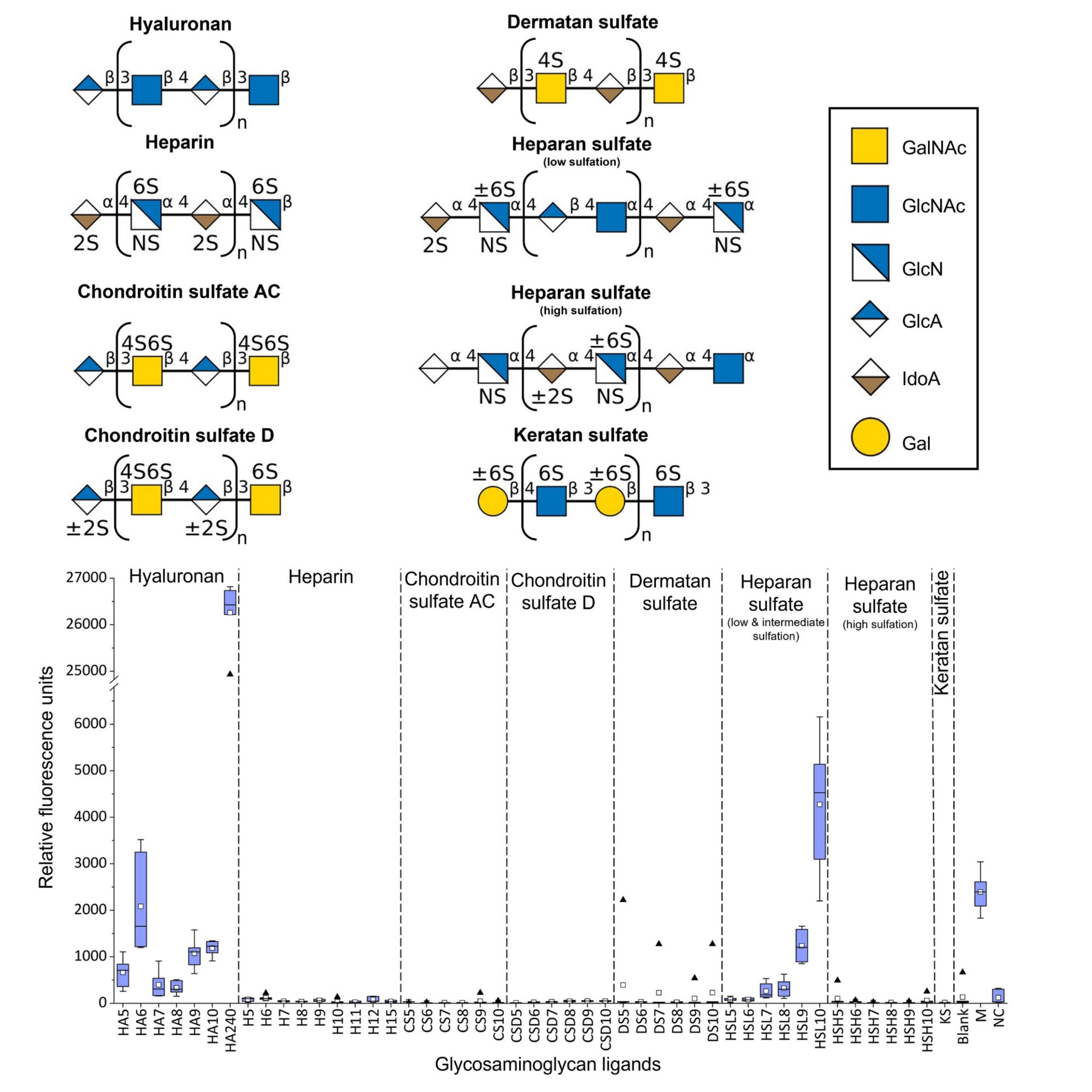
To investigate, the team used the Z Biotech Glycosaminoglycan Array, which presents 47 GAG structures spanning different chain lengths and sulfation levels. The panel includes hyaluronan, heparan sulfate, heparin, chondroitin sulfate, dermatan sulfate and keratan sulfate.
The researchers applied fluorescently labeled XN-IL to the array and measured binding across six replicate spots for each ligand.
The results revealed a clear preference: XN-IL recognized hyaluronan and low-sulfated heparan sulfate, while the other GAG classes tested showed background-level signals.
The array also allowed the team to compare responses across chain lengths and sulfation levels. Among ligands with 10 disaccharide repeats, low-sulfated heparan sulfate produced approximately 3.7 times the fluorescence signal of hyaluronan. The longest hyaluronan tested, HA240, produced the highest overall signal.
These comparisons helped the researchers move beyond identifying a single binding partner to defining a broader recognition profile. They showed that XN-IL did not bind GAGs indiscriminately: the type of GAG and its sulfation level mattered.
Connecting binding specificity to structure
The array established which GAGs XN-IL recognized under the assay conditions. Structural and biophysical studies then helped explain the interaction, revealing how glucuronic acid engages the lectin’s calcium-dependent binding site.
Together, these findings supported the authors’ proposal that XN-IL could serve as a new probe for studying GAGs, with binding that can be reversed by adding EDTA.

Figure 1. Binding of fluorescently labeled XN-IL to 47 GAG structures on the Z Biotech Glycosaminoglycan Array. Each ligand was measured in six replicate spots. XN-IL showed binding to hyaluronan (HA) and lowsulfated heparan sulfate (HSL), with background-level responses for the other GAG classes tested. Numbers following ligand codes indicate the number of disaccharide repeats. M, array marker; NC, printingbuffer negative control. Reproduced from Korsák et al., Carbohydrate Polymers 388, 125557 (2026), under CC BY 4.0.
What this means for your research
A broad glycan screen can reveal a promising interaction, but a dedicated GAG panel can help resolve the specificity behind it. Testing only one GAG, such as heparin, may miss proteins that prefer less-sulfated structures – as this study illustrates.
The Z Biotech Glycosaminoglycan Array allows researchers to:
For the XN-IL study, the array turned an initial hyaluronan hit into a more informative binding profile, helping reveal an unexpected specificity within a familiar lectin family.
Korsák, M., Komárek, J., Houser, J., Pongener, I., Miller, G. J., & Wimmerová, M. (2026). A glycosaminoglycan-binding LecA-like lectin from Xenorhabdus nematophila: structural and biophysical characterization. Carbohydrate Polymers, 388, 125557. https://doi.org/10.1016/j.carbpol.2026.125557